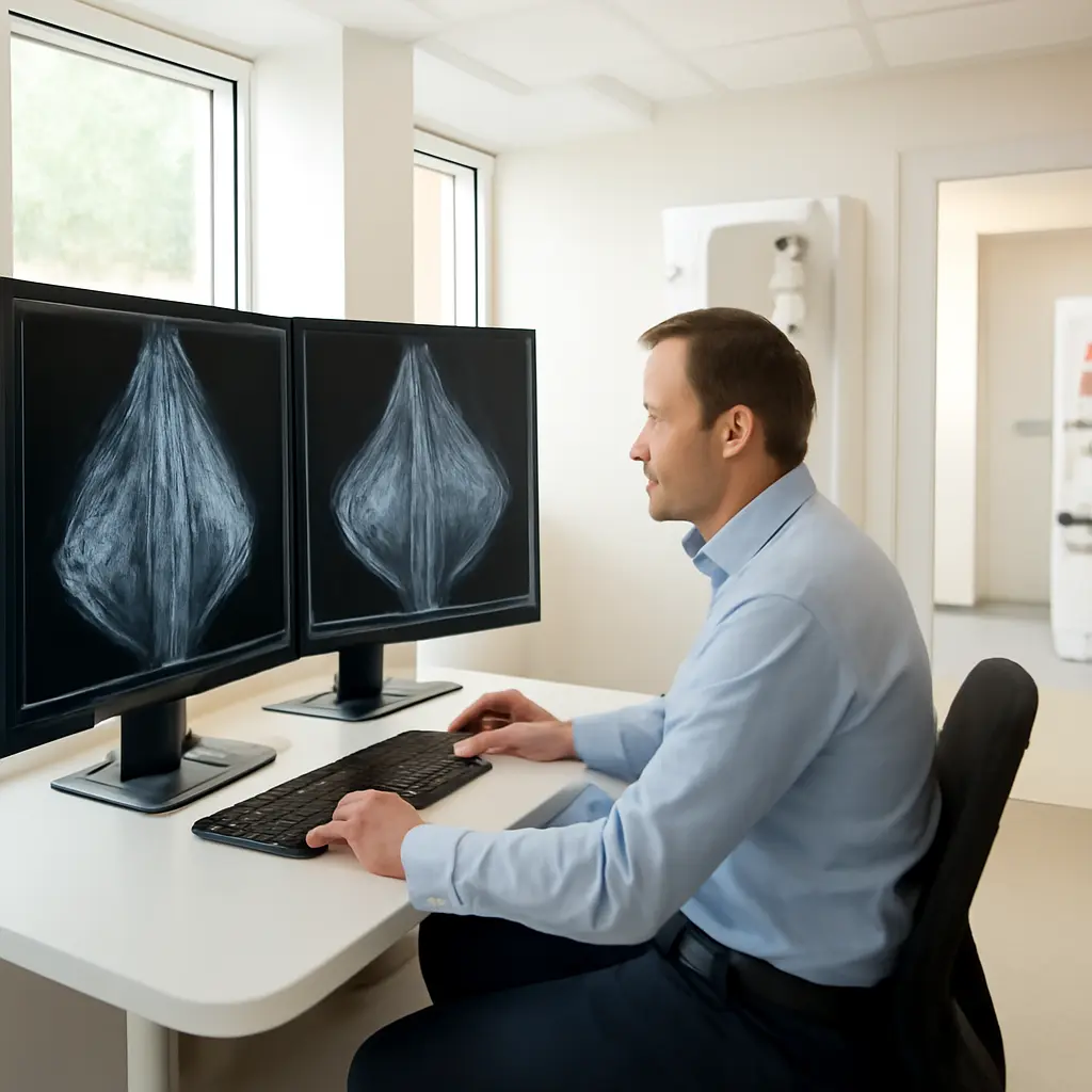
Breast shape changes are differences in the contour, position, or symmetry of breast tissue that may be visible on a mammogram or other breast images. Radiologists look for changes such as a new distortion, a developing asymmetry, or a mass that pulls nearby tissue out of its usual arrangement. These findings can be subtle and are not, by themselves, proof of cancer: normal anatomy, positioning, and benign conditions can produce similar appearances. Their importance lies in comparison with prior images and, when warranted, additional imaging or biopsy. The American College of Radiology’s BI-RADS system standardizes how breast imaging findings are described and managed; the National Cancer Institute estimates that about 1 in 8 women in the United States will develop breast cancer during their lifetime.
Radiologists assess breast shape changes through imaging features
“Breast shape change” is a plain-language description, not a single formal diagnosis. In mammography, radiologists characterize the underlying imaging finding—such as architectural distortion, asymmetry, or a mass—and judge whether it is new, persistent, and suspicious. The American College of Radiology’s Breast Imaging Reporting and Data System (BI-RADS) provides common terminology and assessment categories so findings can be communicated consistently and linked to follow-up recommendations.
A change matters most when it is new or has evolved compared with earlier studies. Radiologists consider the breast’s normal tissue pattern, the image angle and compression, and whether the finding appears on more than one view. A possible abnormality on a screening image may therefore lead to diagnostic mammography, targeted ultrasound, or, in selected cases, MRI rather than an immediate conclusion.
Architectural distortion: tissue appears pulled or rearranged
Architectural distortion is an abnormal arrangement of breast tissue without a clearly defined mass. Fibrous lines may converge, or the normal pattern may look retracted or twisted. The finding can be subtle, particularly in dense tissue, and may become clearer on diagnostic views such as tomosynthesis or spot-compression images.
Distortion can result from benign causes, including prior surgery, trauma, or scarring, but it can also be associated with malignancy. The Radiographics review by Gaur and colleagues describes architectural distortion as an important mammographic finding that may require further evaluation even when no distinct lump is visible. Radiologists use prior images and additional views to distinguish a stable scar from a new or unexplained pattern; if suspicion remains, tissue sampling may be recommended.
Developing asymmetry: a difference that is new or growing
An asymmetry is an area of breast tissue that looks denser or different from the corresponding area in the other breast, but does not meet the imaging definition of a mass. A developing asymmetry is one that is new or more conspicuous on later examinations. That change over time makes it more important to investigate than a stable asymmetry that has appeared unchanged across prior studies.
The finding may reflect normal tissue overlap, hormonal or benign changes, or a lesion that needs assessment. Diagnostic mammographic views help determine whether the apparent difference persists when tissue is repositioned or compressed. Targeted ultrasound may then be used to look for a correlate. The American College of Radiology’s BI-RADS framework guides whether the result calls for routine screening, short-interval follow-up, or biopsy.
Masses and contour changes: shape is considered with other features
A mass is a three-dimensional finding seen on multiple mammographic views. Radiologists describe its shape, margins, and density, then combine those features with its location and change over time. A spiculated or irregular mass can be more concerning than a smoothly circumscribed one, but appearance alone does not establish a diagnosis.
Some breast cancers also cause visible changes in the skin or nipple, including retraction or tethering, while many benign conditions can alter contour as well. Imaging and clinical examination provide complementary information. A new persistent change—especially one that is localized or accompanied by a lump, skin dimpling, or nipple inversion—should be discussed with a healthcare professional rather than judged from appearance alone.
Radiologists validate breast shape changes by comparing images and using targeted tests
The central task is to determine whether a shape difference is real, reproducible, and clinically meaningful. Mammograms compress the breast and show it from different angles, so tissue overlap can mimic an abnormality on one image. Comparison with earlier studies can reveal whether a feature has been stable for years or is newly developing—an essential distinction in breast imaging.
Prior images and multiple views reduce uncertainty
Radiologists review the screening images in standard views and may request additional diagnostic projections. Spot compression spreads overlapping tissue, while tomosynthesis creates thin image slices that can make tissue architecture easier to assess. If the apparent distortion disappears when the breast is repositioned, overlap may have contributed; if it persists, further evaluation may be appropriate.
The U.S. Food and Drug Administration requires mammography facilities to meet quality standards under the Mammography Quality Standards Act. Even with quality-controlled imaging, some findings remain indeterminate until evaluated with additional imaging or biopsy. BI-RADS assessment categories communicate the radiologist’s level of concern and the recommended next step, not a guarantee of a particular outcome.
Ultrasound, MRI, and biopsy answer different questions
Targeted ultrasound can assess a specific area and determine whether it has a visible correlate, such as a cyst or solid lesion. MRI may be used in selected situations—for example, when conventional imaging leaves an important question unresolved or when a person’s overall risk warrants supplemental screening. Neither test replaces mammography for every patient; the appropriate choice depends on the finding and clinical context.
When imaging remains suspicious, a radiologist may recommend image-guided core needle biopsy. Biopsy examines tissue directly and is the way to establish whether a suspicious imaging feature is cancer. A recommendation for biopsy signals that clarification is needed; it does not mean cancer has already been diagnosed.
Breast shape findings need context, not self-diagnosis
Breast tissue varies naturally, and benign conditions, prior procedures, and image positioning can all affect shape. The most useful clues for radiologists are a finding’s precise appearance, whether it persists on additional views, and whether it is new or changing compared with earlier images. This is why attending recommended follow-up matters, even when there is no palpable lump or discomfort.
For patients, the practical step is to keep screening appointments, make prior images available when changing facilities, and follow the report’s recommendation for additional evaluation. Anyone who notices a new persistent breast or nipple change should contact a healthcare professional. Imaging interpretation is individualized; this article is educational and does not replace medical advice.
Conclusion: subtle shape changes are clues that guide careful evaluation
Architectural distortion, developing asymmetry, and changes associated with a mass are different imaging patterns that can alter breast shape or tissue arrangement. Radiologists assess them through multiple views, comparison with prior images, and, when needed, ultrasound, MRI, or biopsy. Because both benign conditions and cancer can produce changes, the finding’s context—not shape alone—determines the next step. Follow the recommendations in your imaging report and ask your care team to explain any requested follow-up.
Sources: American College of Radiology, BI-RADS: Mammography, https://www.acr.org/Clinical-Resources/Reporting-and-Data-Systems/Bi-Rads; National Cancer Institute, Breast Cancer—Patient Version, https://www.cancer.gov/types/breast; Gaur S, et al., “Architectural Distortion of the Breast,” RadioGraphics, 2013, https://doi.org/10.1148/rg.333125176; U.S. Food and Drug Administration, Mammography Quality Standards Act and Program, https://www.fda.gov/radiation-emitting-products/mammography-quality-standards-act-and-program